

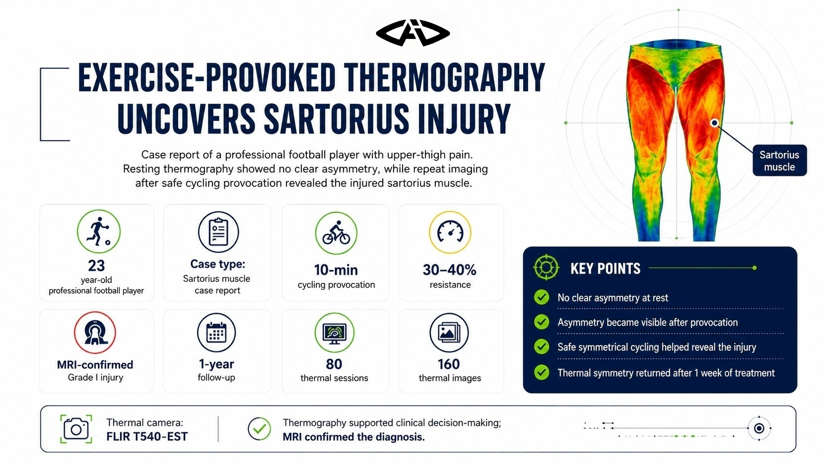
A sartorius muscle injury that was not clearly visible on resting thermography became detectable after repeat thermal imaging following safe, controlled exercise. This case highlights the value of evaluating symptomatic athletes not only at rest, but also after an appropriate provocation protocol.
Case Overview
The study evaluated a 23-year-old professional football player who reported pain in the upper thigh. He experienced pain while lifting the right knee, jogging, and especially while putting on his socks.
The first resting thermographic image did not show a clear right-left asymmetry indicating injury. The researchers therefore applied a controlled exercise protocol designed to safely provoke the suspected sartorius muscle.
Key takeaway: A normal-looking resting thermogram may not exclude a low-grade muscle injury. Repeat imaging after safe exercise can reveal hidden thermal asymmetry.
Imaging and Exercise Protocol
Before imaging, the athlete remained for 5 minutes in a thermally insulated room maintained at 21 °C. Images were obtained in a standard anatomical posture at a fixed distance of 2 metres from the thermal camera.
After the first image, the athlete completed:
- 10 minutes of cycling
- 30–40% resistance
- symmetrical loading of both lower limbs
- supervision by a specialist physiotherapist
Cycling was selected because it could stimulate the sartorius muscle while distributing load equally between both legs. The aim was to observe the muscle response without increasing the severity of a possible injury.
Resting and Post-Exercise Findings
The resting thermogram showed no clear asymmetry that directly identified the injury. However, the player had likely been protecting the painful side during daily activity and placing more load on the healthy leg, which complicated interpretation.
After 10 minutes of cycling, the right sartorius muscle showed a visible temperature increase and a clear asymmetric thermal pattern.
| Assessment | Mean proximal sartorius difference |
|---|---|
| Resting image | 0.36 °C |
| Post-exercise image | 0.64 °C |
After exercise, the reported minimum temperature difference in the proximal region was 1.34 °C, while the maximum temperature difference was 0.77 °C. The provocation protocol made the thermal response of the suspected right sartorius injury more visible.
MRI Confirmation
MRI was performed after the thermographic assessment. The MRI report confirmed a Grade I sartorius muscle injury.
This result showed that the asymmetry revealed after exercise was compatible with the muscle injury. In this case, thermography was used as a supportive early assessment tool rather than as a stand-alone definitive diagnostic method.
Treatment and Follow-Up
Treatment was initiated, and protective kinesiotape was used for one week. Thermal imaging was repeated after the athlete completed a pain-free field training session one week later.
Thermal symmetry had returned in the sartorius regions:
| Region | Mean temperature difference |
|---|---|
| Proximal | 0.06 °C |
| Middle | 0.13 °C |
| Distal | 0.03 °C |
The athlete reported that his pain had disappeared on the same day thermal symmetry was restored. The symmetrical thermal appearance remained present even after 90 minutes of training.
One-Year Monitoring
The athlete was followed weekly before and after training for one year.
- 80 thermal sessions
- 160 anterior and posterior images
- 1 year of follow-up
This long-term monitoring supported the use of thermography not only during the initial injury assessment, but also throughout rehabilitation follow-up.
Interpreting the Case
The case showed that protective loading can make resting thermography difficult to interpret. When the athlete reduces load on the painful side, the healthy side may appear warmer, while the actual injured area remains less obvious at rest.
A safe exercise that loads both sides equally and matches the functional role of the suspected muscle can reveal the injured muscle’s true thermal response.
Clinical note: When a symptomatic athlete shows no clear asymmetry at rest, repeat thermography after a safe, muscle-specific, symmetrical exercise may provide additional information.
Conclusion
This case demonstrates the supportive value of thermography in sartorius injuries, which are rare and may resemble other upper-thigh injuries. Repeat imaging after safe exercise provocation helped reveal a Grade I injury that was not clear on the resting image.
Infrared thermography may contribute to early assessment and rehabilitation monitoring as a fast, non-invasive and practical method. However, findings should be interpreted alongside confirmatory imaging such as MRI.