

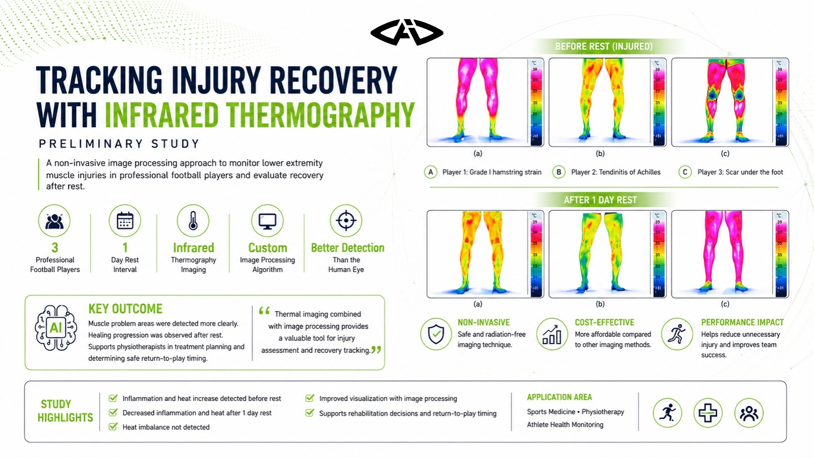
Lower-extremity muscle problems in professional football players were compared before and after rest using infrared thermography and image processing. Across three documented cases, the method made problem areas easier to visualise and showed reduced heat and inflammation after one day of rest.
Study Objective
Early recognition of muscle problems and monitoring recovery are important for rehabilitation planning in football. This preliminary study combined infrared thermography with image processing to support physiotherapists in evaluating lower-extremity muscle problems.
Professional players with documented injuries or muscle-related problems were assessed before rest and again after one day of rest.
Key message: Simplifying thermal images with image processing can make affected muscle regions more visible and help track recovery after rest.
Players Included
Three professional football players from the Turkish Süper Lig level were included.
| Player | Documented problem |
|---|---|
| Player 1 | Grade I hamstring strain |
| Player 2 | Achilles tendinitis |
| Player 3 | Uneven loading related to a scar under the foot |
The players were imaged with infrared thermography after a training match. They were then rested for one day and assessed again.
Image-Processing Method
A custom image-processing approach was used to highlight warmer regions in the thermographic images.
The steps were:
1. Determine the lower and upper RGB values of the thermal colour palette. 2. Threshold areas corresponding to higher temperatures. 3. Create a binary mask for the warmer regions. 4. Convert the original image to grayscale. 5. Blend the coloured warm-region pixels with the grayscale image.
As a result, high-temperature regions remained coloured while the rest of the image appeared in grayscale. The aim was to help physiotherapists inspect problem areas more easily.
Player 1: Hamstring Strain
The first player had a Grade I strain with increased heat and inflammation in the right inner hamstring. After image processing, the location and affected muscle region were easier to distinguish.
Following treatment and one day of rest:
- heat in the right inner hamstring approached normal,
- only a small area remained,
- recovery was visible on the thermal images.
The paper stated that without rest and treatment, the problem could have become more serious and the player might have missed about 20 days of training and competition.
Player 2: Achilles Tendinitis
The second player had Achilles tendinitis associated with increased heat in the right leg. Initial images showed heat on both legs, but image processing indicated that the main problem was located in the right inner calf.
The heat on the left side was linked to compensatory loading because the player protected the painful right leg and used the left leg more. The study described this as reflected heat.
After one day of rest and treatment:
- heat in the right inner calf returned toward normal,
- reflected heat on the left side disappeared,
- recovery was observed.
The article stated that progression of the problem could have required up to 50 days of treatment.
Player 3: Foot Scar and Compensation
The third player had a scar under the right foot. Because of pain, he reduced loading on the right side, which produced increased heat in the left calf.
Thermography and image processing showed not only the scar-related problem but also the altered lower-extremity loading pattern.
After treatment and one day of rest:
- calf heat decreased,
- thermal asymmetry approached normal,
- signs of compensatory loading disappeared.
The study stated that continued exercise without identifying the problem could have caused the player to miss up to 30 days.
Shared Findings After Rest
Across all three players, one day of rest and treatment was followed by:
- reduced inflammation and heat,
- no remaining clear heat imbalance,
- self-reported improvement.
Clinical note: Increased heat on the opposite side does not always indicate a direct injury. Compensatory loading caused by protecting the painful side can also alter thermal distribution.
Interpretation
This preliminary study showed that infrared thermography combined with image processing may support the assessment of muscle problems in professional football players. The algorithm highlighted warmer areas in a form that was easier for clinicians to interpret.
Repeat imaging after one day of rest also made changes in heat and inflammation visible. The method therefore supported both problem localisation and recovery monitoring.
Limitation
This was a preliminary study involving only 3 professional football players. The findings should therefore be validated with larger samples and more advanced image-processing methods.
Conclusion
When infrared thermography and image processing are used together, they may help visualise lower-extremity muscle problems and track recovery more clearly. The approach can provide supportive information for treatment planning, rest decisions and the timing of rehabilitation completion.